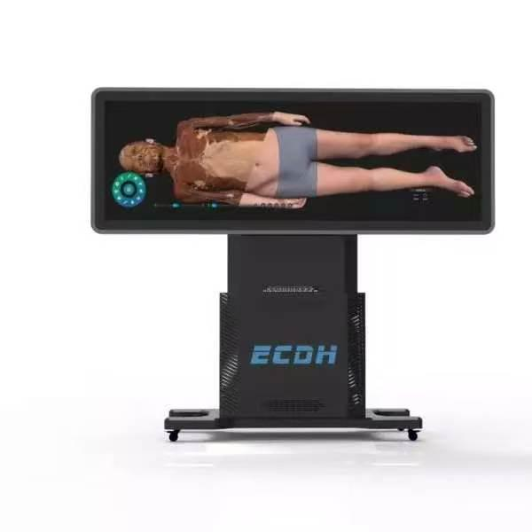
The landscape of medical education is shifting rapidly. Educators now balance the timeless necessity of gross anatomy with the need for modern, repeatable, and non-toxic training environments. As institutions move away from traditional cadaver-only models, digital innovation provides a clear path forward for surgical training and anatomical study.
Bridging the Gap in Modern Anatomy Training
For decades, the standard for learning human structure involved formaldehyde-based preservation. While useful, this approach poses significant health risks and financial burdens to universities and hospitals. Maintaining a lab requires ongoing costs for cadaver procurement, specialized ventilation, and strict regulatory compliance.
The integration of advanced digital tools has changed how students visualize complex systems. Educators can now move beyond static illustrations or one-time dissection sessions. A high-performance virtual anatomy table allows instructors to demonstrate intricate vascular networks, muscular layers, and organ systems with repeatable digital practice. Students can repeat virtual dissections as needed without affecting the digital model.
Precision and Data-Driven Learning
DIGIHUMAN has engineered its technology to meet the rigorous demands of professional medical study. The core innovation lies in the extreme level of detail provided by its imaging data. The system utilizes real human anatomical imaging datasets, featuring reconstructed cross-sections with a layer thickness as thin as 0.1mm. This level of resolution provides a detailed view of human morphology.
The software platform is built on continuous cross-sectional datasets derived from real human specimens. This comprehensive repository of visual information ensures that users explore actual human structure rather than simplified artistic renders. With over 6,000 reconstructed anatomical structures spanning key biological systems, these structures preserve detailed anatomical relationships based on the source datasets.
This level of precision is essential for pre-operative planning and academic research. When clinicians can zoom into a specific nerve bundle or trace the exact path of an artery through multiple planes, the educational value far exceeds that of a textbook.
Scalability and Implementation
One of the primary advantages of this virtual anatomy table is its versatility within a facility. Unlike a physical dissection lab that occupies large amounts of floor space and specialized infrastructure, the digital unit is compact and easy to deploy across classrooms, lecture halls, or surgical simulation centers.
The software platform also bridges the gap between different diagnostic tools. Because the data is based on high-resolution imaging, it aligns seamlessly with modern clinical workflows. It allows medical professionals to correlate anatomical structures with CT and MRI scans. This integration is vital for radiology students and surgeons who must interpret cross-sectional imaging in their daily practice.
Furthermore, the technology supports a variety of interactive features. Users can toggle layers on or off to isolate specific body systems. They can also create cross-sectional views at any angle, providing a multidimensional understanding that was previously difficult to achieve without destructive methods.
Interactive Assessment and Multi-User Collaboration
Modern medical education relies heavily on active, collaborative learning environments. The system supports multi-touch interactivity, allowing small groups of students to work simultaneously on complex anatomical cases. Instructors can assign interactive tasks, slice through organ models together, and analyze clinical anomalies in real time.
To reinforce knowledge retention, the platform includes built-in assessment and self-study modules. Educators can create customized quizzes, pin specific structures for identification exams, and track individual student progress over time. This structured approach helps bridge the gap between theoretical study and practical board exam preparation.
Furthermore, integrating VR and AR technologies alongside 3D medical models provides students with immersive spatial orientation. Learner engagement increases significantly when students can manipulate anatomical structures in full three-dimensional space.
The Role of DIGIHUMAN in Future Surgical Training
As the medical field continues to embrace digitalization, the demand for high-fidelity simulation tools will only grow. The virtual anatomy table represents a cornerstone of this transition. It is an additional option alongside cadaver-based anatomy education while simultaneously expanding access to high-quality anatomical education.
The system is designed with a user-friendly interface, ensuring that the focus remains on the learning objectives rather than navigating complex software. By digitizing the human body with such high accuracy, institutions can ensure that every student, regardless of their specialization, has access to consistent, high-level anatomical training.
For university administrators, DIGIHUMAN may help optimize long-term laboratory operations. It reduces the dependency on external supply chains for biological materials and creates a modular space that can adapt to different curricula. Whether the goal is to enhance a basic undergraduate anatomy course or to provide advanced surgical planning tools for hospital residents, the data-driven approach provides a reliable, robust solution.
Modern medical schools are moving toward a blended learning environment. This hybrid approach combines the irreplaceable experience of gross anatomy with the precision and convenience of digital platforms. By leveraging these tools, institutions ensure that the next generation of physicians is not only prepared for the operating room but is also proficient in interpreting the complex, multidimensional data that defines modern medicine.